|
|
||
From:
Hélène Berthoumieux1,2,3, Jean-Léon Maître4,5, Carl-Philipp Heisenberg4, Ewa K Paluch6, Frank Jülicher1 and Guillaume Salbreux1
1 Max Planck Institute for the Physics of Complex Systems, Nöthnitzerstr. 38, 01187 Dresden, Germany 2 Sorbonne Universités, UPMC Univ Paris 06, UMR 7600, LPTMC, F-75005, Paris, France 3 CNRS, UMR 7600, LPTMC, F-75005, Paris, France
4 Institute of Science and Technology Austria, Klosterneuburg, Austria 5 EMBL, Meyerhofstrasse 1, 69117 Heidelberg, Germany 6 MRC LMCB, University College London, Gower Street, WC1E 6BT, London, UK E-mail: salbreux@pks.mpg.de
“Active elastic thin shell theory for cellular deformations”, New Journal of Physics, Vol 16, 2014 065005,
DOI: 10.1088/1367-2630/16/6/065005
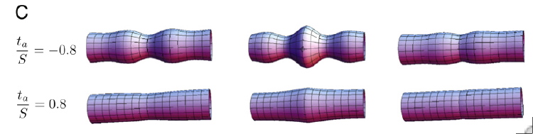
ABSTRACT: We derive the equations for a thin, axisymmetric elastic shell subjected to an internal active stress giving rise to active tension and moments within the shell. We discuss the stability of a cylindrical elastic shell and its response to a localized change in internal active stress. This description is relevant to describe the cellular actomyosin cortex, a thin shell at the cell surface behaving elastically at a short timescale and subjected to active internal forces arising from myosin molecular motor activity. We show that the recent observations of cell deformation following detachment of adherent cells (Maître J-L et al 2012 Science 338 253–6) are well accounted for by this mechanical description. The actin cortex elastic and bending moduli can be obtained from a quantitative analysis of cell shapes observed in these experiments. Our approach thus provides a non-invasive, imaging-based method for the extraction of cellular physical parameters.
This is Fig. 3C from the paper:
3D representation of the SSSS
cylinder deformation induced by a localized increase of active tension or moments.
Page 169 / 360