|
|
||
From:
Hélène Berthoumieux1,2,3, Jean-Léon Maître4,5, Carl-Philipp Heisenberg4, Ewa K Paluch6, Frank Jülicher1 and Guillaume Salbreux1
1 Max Planck Institute for the Physics of Complex Systems, Nöthnitzerstr. 38, 01187 Dresden, Germany 2 Sorbonne Universités, UPMC Univ Paris 06, UMR 7600, LPTMC, F-75005, Paris, France 3 CNRS, UMR 7600, LPTMC, F-75005, Paris, France
4 Institute of Science and Technology Austria, Klosterneuburg, Austria 5 EMBL, Meyerhofstrasse 1, 69117 Heidelberg, Germany 6 MRC LMCB, University College London, Gower Street, WC1E 6BT, London, UK E-mail: salbreux@pks.mpg.de
“Active elastic thin shell theory for cellular deformations”, New Journal of Physics, Vol 16, 2014 065005,
DOI: 10.1088/1367-2630/16/6/065005
ABSTRACT: We derive the equations for a thin, axisymmetric elastic shell subjected to an internal active stress giving rise to active tension and moments within the shell. We discuss the stability of a cylindrical elastic shell and its response to a localized change in internal active stress. This description is relevant to describe the cellular actomyosin cortex, a thin shell at the cell surface behaving elastically at a short timescale and subjected to active internal forces arising from myosin molecular motor activity. We show that the recent observations of cell deformation following detachment of adherent cells (Maître J-L et al 2012 Science 338 253–6) are well accounted for by this mechanical description. The actin cortex elastic and bending moduli can be obtained from a quantitative analysis of cell shapes observed in these experiments. Our approach thus provides a non-invasive, imaging-based method for the extraction of cellular physical parameters.
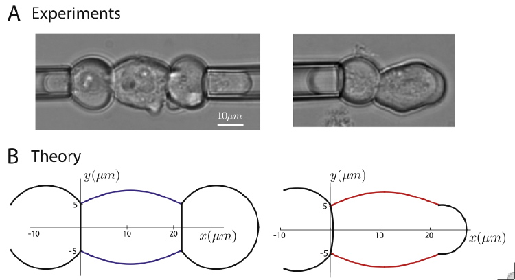
This is Figure 5 from the paper:
Figure 5. Experimental and theoretical cell deformation before and after cell detachment. The theoretical cell shape is calculated using parameters obtained from a fit to experimental measurements of cell deformation. (A) Cell triplet visualized with transmission microscopy before and after cell detachment. (B) Initial and final shapes of the cell triplet obtained using active elastic shell theory. The initial shape of the middle cell body is obtained by solving equation (24). The two side cells have spherical cap shapes. The interfaces are flat. The final shape of the middle cell body is obtained by solving equations (28)–(30) with boundary conditions given in equations (38)–(45). The side cell, interface, and bulge have a spherical cap shape. The values of geometrical parameters used to determine these shapes are given in tables 1 and 2.
Page 170 / 360