|
|
||
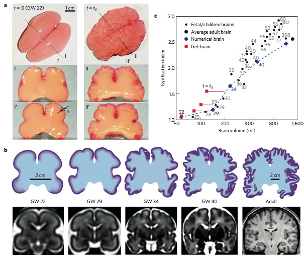
Figure 2 | Sectional views of model brains during convolutional development. a, Planform and cross-sectional images of a physical gel-brain showing convolutional development during the swelling (folding) process that starts from an initially smooth shape (left panels) to a moderately convoluted shape (right panels). b, The coronal sections of the simulated brain (top panels) with comparisons to corresponding MRI sections. c, Gyrification index as a function of brain size for real brains (data from refs 3,40), a numerically simulated brain, and a physical gel-brain. Gestational week is indicated for fetal and children brains. The initial volume of the gel-brain (≈34 ml) is scaled to match that of the simulated brain (≈57 ml). Note that in the gel experiments only the outer layer swells and therefore the volume grows less than in real brains.
From:
Tallinen T., Chung J., Rausseau F., Girard N., Lefevre J., Mahadevan L. “On the growth and form of cortical convolutions”, Nat. Phys., 12 (2016), pp. 588-593
ABSTRACT: The rapid growth of the human cortex during development is accompanied by the folding of the brain into a highly convoluted structure. Recent studies have focused on the genetic and cellular regulation of cortical growth, but understanding the formation of the gyral and sulcal convolutions also requires consideration of the geometry and physical shaping of the growing brain. To study this, we use magnetic resonance images to build a 3D-printed layered gel mimic of the developing smooth fetal brain; when immersed in a solvent, the outer layer swells relative to the core, mimicking cortical growth. This relative growth puts the outer layer into mechanical compression and leads to sulci and gyri similar to those in fetal brains. Starting with the same initial geometry, we also build numerical simulations of the brain modelled as a soft tissue with a growing cortex, and show that this also produces the characteristic patterns of convolutions over a realistic developmental course. All together, our results show that although many molecular determinants control the tangential expansion of the cortex, the size, shape, placement and orientation of the folds arise through iterations and variations of an elementary mechanical instability modulated by early fetal brain geometry.
Page 79 / 360